| PREVIOUS PRESENTATION | BACK TO PROGRAM OVERVIEW | NEXT PRESENTATION |
Subcycle optical spectroscopy at the atomic scale
Tom Siday
School of Physics and Astronomy, University of Birmingham, Birmingham, United Kingdom
Measuring the interaction of light and matter over the shortest possible length- and timescales has been a long-sought goal in condensed matter physics. By exploiting evanescent fields confined to miniscule objects, near-field micro¬scopy can access ultrafast light-matter interaction on nanometer length scales [1]. Yet, the spatial resolution accessible to near-field microscopy is strictly limited by the size of the probe apex (~10 nm). Tantalizing glimpses into even smaller spatial scales became possible with the advent of lightwave-driven scanning tunnelling microscopy (LW-STM) [2],[3]. However, by sampling only the time-integrated electronic response using rectified currents, the rapid motion of electrons within a single cycle of light remains out of reach. Here, we demonstrate a fundamentally new paradigm for optical microscopy (Fig. 1a), which exploits atomic nonlinearities within optical near-fields to achieve atomic-scale spatial resolution and subcycle time resolution [4].
To explore the role of atomic protrusions in near-field microscopy, we use qPlus atomic force microscopy sensors under ultra-high vacuum and cryogenic conditions. We illuminate the tip apex with strong, phase-locked terahertz (THz) pulses, and detect scattered light with electro-optical sampling (EOS). For tip-sample separations larger than ~1 nm, we observe a conventional near-field response (Fig. 1b, blue lines). Intriguingly, when the tip-sample separation reaches the atomic scale, the signal dramatically increases, accruing a phase delay Δφ. This emergent signal decays at the same rate as the time-integrated tunnelling currents 〈Jlw 〉 in LW-STM (Fig. 1e), confirming its origin: electromagnetic radiation emitted by tunnelling currents flowing in response to the THz electric field (Fig. 1c,d). We confirm this using ab initio quantum simulations, and demonstrate how near-field optical tunnelling emission (NOTE) can image single packing defects on the surface of Au(111) (Fig. 1f). Finally, we sample the real-time quantum flow of electrons traversing a WSe2 trilayer, without requiring any a priori assumptions on tunnelling (Fig. 1g). The subcycle optical sampling at the heart of NOTE provides experimental access to atomic-scale electron dynamics in a broad range of quantum materials – even insulators. This widely tuneable all-optical approach opens the door to strong-field control over light-matter interaction on atomic length scales.
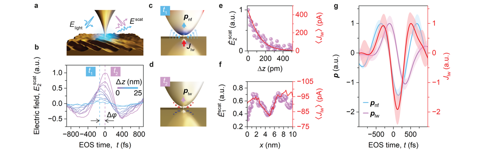
Figure 1: NOTE microscopy. (a) A THz light pulse (Elight) is coupled to a tungsten tip tapping near the surface of Au(111), with scattered near-fields (Escat) detected using EOS. (b) Scattered THz transients (E2scat) for increasing tip-sample separation Δz (tip tapping amplitude A = 25 nm). For minimal Δz, the transient transforms, accruing a phase shift Δφ, and a dramatic amplitude increase relative to the conventional near fields. (c) Formation of a mesoscopic near-field dipole pnf at the tip apex, driven by Elight at time t1 where Elight is maximal, causing a tunnelling current Jlw to flow. (d) At time t2, pnf = 0 as Elight crosses zero, but the tunnelling current induced NOTE dipole plw is at a maximum. (e) Peak of the NOTE signal E2scat for A = 200 pm, alongside the time-integrated lightwave tunnelling current 〈Jlw〉 measured for increasing Δz. (f) Quasi-constant height LW-STM and NOTE line scan measured across a single packing defect on the surface of Au(111). (g) Real-time ultrafast tunnelling currents sampled on trilayer WSe2, showing pnf at the tip apex (blue), the NOTE dipole plw (purple) and the retrieved ultrafast tunnelling currents (red).
References
[1] M. Plankl et al., “Subcycle contact-free nanoscopy of ultrafast interlayer transport in atomically thin heterostructures”, Nat. Photonics 15, 594 (2021)
[2] T. L. Cocker et al., “An ultrafast terahertz scanning tunnelling microscope”, Nat. Photonics 7, 620 (2013)
[3] T. L. Cocker et al., “Tracking the ultrafast motion of a single molecule by femtosecond orbital imaging”, Nature 539, 263 (2016)
[4] T. Siday et al., “All-optical subcycle microscopy on atomic length scales”, Nature 629, 329 (2024)